e- Cyborg Plants with using electronic circuits for trees in the garden and in the park AMNIMARJESLOW GOVERNMENT 91220017 XI XAM PIN PING HUNG CHOP 02096010014 LJBUSAF e- cyborg tree for substitute reliability, beauty and durability of plants ___ Thankyume ON Lord Jesus Blessing predicate beautiful light in the garden and park ___ JESS Circular growing electronics and PIT Instrument to Count Rolling ___ Gen. Mac Tech Zone e- Cyborg Tree on Electronics tree
when the advancement of electronic technology continues to grow where the growth of cells in plants can be compared and replaced and combined with a dynamic electron motion system, we can make various types of plants that can grow, flower and bud grow using robotic electronics techniques. here I will describe the workings and possible solutions to environmental impacts and aspects of their wisdom for natural living beings.
Wired and weird: Meet the cyborg plants
From electrified roses to robotic roots, engineers are taking plants high-tech
Tiny technology gives plants a boost
The cyborg leaves of this weedy mustard relative contain tiny antennae. They help the plant capture more light, making more energy than normal leaves do.
Michael Strano, MIT
Two years before the Linköping group published its findings, Strano’s team made an exciting discovery. This MIT group found a way to insert extremely tiny machines — nano-machines — into a plant’s chloroplasts. These researchers work in a branch of engineering known as nanotechnology. They study incredibly tiny materials that that operate at very small scales.
Before Strano’s research, scientists had not found a way to get anything through the wall of a plant cell’s chloroplasts. But the MIT team discovered that when it coated tiny particles with electrically charged molecules, chloroplants would suck those particles straight inside. Strano's team now understood how to insert any tiny particle, material, or even nanomachine into a plant cell.
At last they could begin testing different materials with the goal of altering a plant’s function, such as harvesting sunlight better.
A radio without its antenna can’t capture very clear signals. Similarly, chloroplasts in a regular leaf cannot absorb much of the sun’s light. Plants focus on hues from deep blue to red, ignoring other wavelengths in sunlight. Strano’s team decided to test the creation of a nano-antenna to boost the range of light a plant would capture. Then they inserted it into the chloroplasts of a small flowering weed within the mustard family.
The nano-antenna likely became trapped in the section of the chloroplast that gathers light, the researchers say. And although they still aren’t yet sure exactly how it works, cyborg plants hosting these antennae produced 30 percent more energy from sunlight than normal.
Under infrared light, parts of this cyborg leaf turn orange. Its orange spots reveal where the tiny antennae have infiltrated the leaf’s cells.
Michael Strano, MIT
The team published its initial findings three years ago, in Nature Materials. Since then, they’ve begun working to further enhance their plants. For instance, they’ve developed tiny sensors that can detect pollutants and other dangers in groundwater. They also found a way to insert these sensors into living plants.
“A plant has roots that extend into the soil. It is constantly drinking,” Strano says. Any pollutants in the soil or groundwater will get pulled in the way the polymers had been in Stavrinidou’s plants. Sensors within a plant’s roots or xylem should then be able to scout for particular chemicals.
“We made an explosive-detecting plant,” Strano says. Here, the plant’s sensors watched for chemicals in water that betrayed the presence of explosives such as TNT. When the sensors detected a bomb, the plant emitted infrared light. To watch for that signal, The MIT team built a system composed of a camera and a computer. When the camera "saw" the infrared light, the computer emailed out a warning.
Robotic roots
Plant roots inspire scientists in other ways, too. They’re remarkably good at finding water and nutrients while avoiding rocks and other obstacles. These are traits that could be especially useful to robots.
The Plantoid robot has a computer inside its trunk that controls each of its thick roots.
Italian Institute of Technology
Roots change direction when they run into objects in the soil. Most roots also are covered in sensitive hairs that check for water and nutrients. That’s how they know to grow toward the nutrients a plant needs. Figuring out how they do this could help researchers build better robots.
That’s why roots caught the attention of Barbara Mazzolai. She is a robot specialist at the Italian Institute of Technology in Genoa. Her team has created a robotic plant and begun running it through its paces. Called Plantoid, it has plastic branches with clear leaves. Each leaf contains sensors that measure temperature, touch and other factors. It has trunk, just like a tree. And this system not only can build its own roots but also direct them where to grow.
The tip of each robo-root uses sensors to gather information from its environment. Those sensors measure levels of nearby nutrients, water and pressure. In response, these roots bend away from obstacles and toward nutrients or water.
Some of the roots can even grow. They extend using a process similar to 3D printing. Plantoid feeds plastic thread from a spool through to the root. It stops at a spot just behind the root’s tip. There, the plastic lays down new layers. Over time, this lengthens the root, pushing the tip out and down. A central computer located in Plantoid’s trunk manages the direction of that growth, based on incoming data from its sensor.
Plantoid has several possible uses. When loaded with sensors, its leaves and roots could spy signs of chemicals or pollutants in the air and soil. Plantoid could even travel to other planets and relay back data about alien worlds.
Lessons learned from building plantoid might one day even lead to new tools in medicine, Mazzolai says. A flexible robot that can lengthen itself might help surgeons enter and operate on difficult-to-reach places. For example, the robot might snake down through the mouth and into the stomach.
Nature electrified
For now, robo-roots and cyborg flowers remain laboratory curiosities. But that may change, because farmers and others are curious about what’s going on in the immediate neighborhood of their crops.
Plantoid’s root pushes through a tub of plastic beads. Similar to a 3D printer, a spool feeds plastic down to just above the root’s tip. This lengthens the root, making it “grow.”
Italian Institute of Technology
Already many farmers use sensors to monitor crops. These are not part of the plants, but instead are external electronic devices set up at regular intervals across a field or greenhouse. These sensors monitor the air or ground for water, nutrients and other things that matter to plant health. When there’s a problem — dry soil, for example — the sensor sends a wireless alert to a central system. Such a system then alerts farmers about exactly what kind of care their crops need.
Cyborg plants might one day interact with or even replace such sensor systems. Stavrinidou’s wires, Strano’s nanomachines, Plantoid and other similar technologies all turn plants into strange new versions of themselves. Cyborg plants could turn out to be healthier, more powerful or even smarter than regular plants.
One day, people may find it normal to walk past roses that both beautify the street and scan for pollution. At the same time, robots inspired by plants may help explore the surfaces of other planets.
Many people head out into the woods or a garden to get away from technology. But one day soon, nature itself may join our connected, electrified world.
Power Words
alien A non-native organism. (in astronomy) Life on or from a distant world.
antenna (plural: antennae) In physics: a device for picking up (receiving) electromagnetic energy.
broadcast To cast — or send out — something over a relatively large distance. A farmer may broadcast seeds by flinging them by hand over a large area. A loudspeaker may send sounds out over a great distance. An electronic transmitter may emit electromagnetic signals over the air to a distant radio, television or other receiving device. And a newscaster can broadcast details of events to listeners across a large area, even the world.
carbon The chemical element having the atomic number 6. It is the physical basis of all life on Earth. Carbon exists freely as graphite and diamond. It is an important part of coal, limestone and petroleum, and is capable of self-bonding, chemically, to form an enormous number of chemically, biologically and commercially important molecules.
carbon dioxide (or CO2) A colorless, odorless gas produced by all animals when the oxygen they inhale reacts with the carbon-rich foods that they’ve eaten. Carbon dioxide also is released when organic matter (including fossil fuels like oil or gas) is burned. Carbon dioxide acts as a greenhouse gas, trapping heat in Earth’s atmosphere. Plants convert carbon dioxide into oxygen during photosynthesis, the process they use to make their own food.
cavity (in geology or physics) A large open region surrounded by tissues (in living organisms) or some rigid pocketlike structure. cell The smallest structural and functional unit of an organism. Typically too small to see with the naked eye, it consists of watery fluid surrounded by a membrane or wall. Animals are made of anywhere from thousands to trillions of cells, depending on their size. Some organisms, such as yeasts, molds, bacteria and some algae, are composed of only one cell.
chemical A substance formed from two or more atoms that unite (become bonded together) in a fixed proportion and structure. For example, water is a chemical made of two hydrogen atoms bonded to one oxygen atom. Its chemical symbol is H2O. Chemical can also be an adjective that describes properties of materials that are the result of various reactions between different compounds.
chlorophyll Any of several green pigments found in plants that perform photosynthesis — creating sugars (foods) from carbon dioxide and water.
chloroplast A tiny structure in the cells of green algae and green plants that contain chlorophyll and creates glucose through photosynthesis.
circuit A network of that transmits electrical signals. In the body, nerve cells create circuits that relay electrical signals to the brain. In electronics, wires typically route those signals to activate some mechanical, computational or other function.
computer chip (also integrated circuit) The computer component that processes and stores information.
computer program A set of instructions that a computer uses to perform some analysis or computation. The writing of these instructions is known as computer programming.
current (in electricity) The flow of electricity or the amount of electricity moving through some point over a particular period of time.
cyborg A living organism that has been given with electronic enhancements.
develop (as with towns) The conversion of wildland to host communities of people. This development can include the building of roads, homes, stores, schools and more. Usually, trees and grasslands are cut down and replaced with structures or landscaped yards and parks.
digital (in computer science and engineering) An adjective indicating that something has been developed numerically on a computer or on some other electronic device, based on a binary system (where all numbers are displayed using a series of only zeros and ones).
electric current A flow of electric charge, called electricity, usually from the movement of negatively charged particles, called electrons.
electricity A flow of charge, usually from the movement of negatively charged particles, called electrons.
electromagnetic The science of sounds and hearing.
electron A negatively charged particle, usually found orbiting the outer regions of an atom; also, the carrier of electricity within solids.
electronics Devices that are powered by electricity but whose properties are controlled by the semiconductors or other circuitry that channel or gate the movement of electric charges.
engineer A person who uses science to solve problems. As a verb, to engineer means to design a device, material or process that will solve some problem or unmet need.
environment The sum of all of the things that exist around some organism or the process and the condition those things create for that organism or process. Environment may refer to the weather and ecosystem in which some animal lives, or, perhaps, the temperature, humidity and placement of components in some electronics system or product.
groundwater Water that is held underground in the soil or in pores and crevices in rock.
infrared light A type of electromagnetic radiation invisible to the human eye. The name incorporates a Latin term and means “below red.” Infrared light has wavelengths longer than those visible to humans. Other invisible wavelengths include X-rays, radio waves and microwaves. It tends to record a heat signature of an object or environment.
internet An electronic communications network. It allows computers anywhere in the world to link into other networks to find information, download files and share data (including pictures).
monitor To test, sample or watch something, especially on a regular or ongoing basis.
nano A prefix indicating a billionth. In the metric system of measurements, it’s often used as an abbreviation to refer to objects that are a billionth of a meter long or in diameter.
nanotechnology Science, technology and engineering that deals with things and phenomena at the scale of a few billionths of a meter or less.
nutrient A vitamin, mineral, fat, carbohydrate or protein that a plant, animal or other organism requires as part of its food in order to survive.
photosynthesis (verb: photosynthesize) The process by which green plants and some other organisms use sunlight to produce foods from carbon dioxide and water.
pigment A material, like the natural colorings in skin, that alter the light reflected off of an object or transmitted through it. The overall color of a pigment typically depends on which wavelengths of visible light it absorbs and which ones it reflects. For example, a red pigment tends to reflect red wavelengths of light very well and typically absorbs other colors. Pigment also is the term for chemicals that manufacturers use to tint paint.
pixelate To break a patterned image up into discrete small boxes, or pixels, of a single color. If the pixels are relatively large, they can make the pattern relatively hard to identify by essentially blurring or erasing small details.
plastic Any of a series of materials that are easily deformable; or synthetic materials that have been made from polymers (long strings of some building-block molecule) that tend to be lightweight, inexpensive and resistant to degradation.
pollutant A substance that taints something — such as the air, water, our bodies or products. Some pollutants are chemicals, such as pesticides. Others may be radiation, including excess heat or light. Even weeds and other invasive species can be considered a type of biological pollution.
polymer A substance made from long chains of repeating groups of atoms. Manufactured polymers include nylon, polyvinyl chloride (better known as PVC) and many types of plastics. Natural polymers include rubber, silk and cellulose (found in plants and used to make paper, for example).
pore A tiny hole in a surface. On the skin, substances such as oil, water and sweat pass through these openings.
power plant An industrial facility for generating electricity.
radio To send and receive radio waves; or the device that receives these transmissions.
robot A machine that can sense its environment, process information and respond with specific actions. Some robots can act without any human input, while others are guided by a human.
sensor A device that picks up information on physical or chemical conditions — such as temperature, barometric pressure, salinity, humidity, pH, light intensity or radiation — and stores or broadcasts that information. Scientists and engineers often rely on sensors to inform them of conditions that may change over time or that exist far from where a researcher can measure them directly.
silicon A nonmetal, semiconducting element used in making electronic circuits. Pure silicon exists in a shiny, dark-gray crystalline form and as a shapeless powder.
three-dimensional (3-D)printing The creation of a three-dimensional object with a machine that follows instructions from a computer program. The computer tells the printer where to lay down successive layers of some raw material, which can be plastic, metals, food or even living cells. 3-D printing is also called additive manufacturing.
transistor A device that can act like a switch for electrical signals.
viable (in biology) Able to survive and/or live a normal lifespan.
wavelength The distance between one peak and the next in a series of waves, or the distance between one trough and the next. Visible light — which, like all electromagnetic radiation, travels in waves — includes wavelengths between about 380 nanometers (violet) and about 740 nanometers (red). Radiation with wavelengths shorter than visible light includes gamma rays, X-rays and ultraviolet light. Longer-wavelength radiation includes infrared light, microwaves and radio waves.
weed (in botany) A plant growing wild in, around — and sometimes smothering over — valued plants, such as crops or landscape species (such as lawn grasses, flowers and shrubs). Often a plant becomes such a botanical bully when it enters a new environment with no natural predators or controlling conditions (such as hard frosts). (in biology, generally) Even an animal may be referred to as a “weed” if it enters an environment and begins to overtake the local ecosystem.
xylem The part of a plant that conducts water, nutrients and sap.
XO__XO Biosensors and Bioelectronics
Biosensors & Bioelectronics is the principal international journal devoted to research, design, development and application of biosensors and bioelectronics. It is an interdisciplinary journal serving professionals with an interest in the exploitation of biological materials in novel diagnostic and electronic devices. Biosensors are defined as analytical devices incorporating a biological material (e.g. tissue, microorganisms, organelles, cell receptors, enzymes, antibodies, nucleic acids etc.), a biologically derived material or a biomimic intimately associated with or integrated within a physicochemical transducer or transducing microsystem, which may be optical, electrochemical, thermometric, piezoelectric or magnetic. Biosensors usually yield a digital electronic signal which is proportional to the concentration of a specific analyte or group of analytes. While the signal may in principle be continuous, devices can be configured to yield single measurements to meet specific market requirements. Biosensors have been applied to a wide variety of analytical problems including in medicine, the environment, food, process industries, security and defence. The emerging field of Bioelectronics seeks to exploit biology in conjuction with electronics in a wider context encompassing, for example, biomaterials for information processing, information storage and actuators. A key aspect is the interface between biological materials and electronics. While endeavouring to maintain coherence in the scope of the journal, the editors will accept reviews and papers of obvious relevance to the community, which describe important new concepts, underpin understanding of the field or provide important insights into the practical application of biosensors and bioelectronics.
BIOSENSORS and BIOELECTRONICS
the areas of biosensors, bioactuators and bioelectronics from bench to bedside. Fundamentals on biosensor design and manufacturing will be covered and translated into clinical applications. In addition, the Workshop will cover the latest advances in biosensors and bioelectronics and related sectors such as mobile and digital health expert systems and distributed diagnostics, and will provide an interdisciplinary workshop for researchers, engineers, clinicians, educators and people from industry to present and discuss the most recent trends and practical challenges encountered in the field of Biosensors and Bioelectronics.
Fundamentals from Materials to Biology and Medicine
The interface of organic electronic materials with cells, neurons, drug actuation
Electrons and ions at the interface with Biology
Active Nanosystems from bench to bedside
( Nanobiosensors, nanomaterials & nanoanalytical systems for diagnosis and therapy of diseases)
Organic and Hybrid nanomaterials
Stimuli-responsive materials
Biosensor design and manufacturing
Microfluidics
Biomolecular Dynamics
Biosensing at biological interfaces
Biomechatronics
Biosensors and Bioactuators
Immuno Sensors
Photonic Sensors and Chemical Sensors
DNA chips & Nucleic Acid Sensors
Enzyme-based Biosensors
Lab-on-a-chip, Organism and Whole Cell-based Biosensors
Biomaterials and Biosensors
Electrodes and transistors
Drug Delivery devices
Neural interfaces
Printed biosensors and Electronic noses
Biological and Clinical Applications
Organic Bioelectronics
Bioelectronics for neurodegenerative, cardiovascular, orthopaedic and other diseases
Biosensors - manufacturing and markets
Biosensors in Drug Delivery
Bioelectronics for Tissue Regeneration
Printed Electronics & Bioelectronics Network for medical devices, implants
Brain/Machine interfaces and prostheses
Bioelectronics, Biocomputing and Biofuel cells
ICT for Wireless Biomedical Sensor Applications
Digital & Mobile diagnostics and Tele-medicine
Computational Modeling
As material systems and device structures become nanosized and nanostructured, significant challenges arise with respect to their design and with respect to tailoring their properties and response in a controlled way. The aim of this Workshop is to explore the state-of-the-art methodologies and approaches for the modelling and optimization of novel materials, the material's behavior and/or nano-device manufacturing processes, as well as multiscale computational approaches that can play a key role towards the further advancement of the corresponding nanotechnologies and enable the manufacturing and characterization of breakthrough devices and systems.
During the Workshop a series of Invited talks, Oral and Poster presentations will be given covering different computational approaches (ranging from the atomic to the macro-scale or multiscale) applied on a broad range of subjects.
Computational approaches to the following :
Material properties and processes at the nano-scale
Computer aided design of novel materials
Structural and mechanical properties
Diffusion and film growth
Photonics, plasmonics, phononics and electronics
Strongly correlated electron systems, magnetism and spintronics
Multiscale modeling of devices and processes
Graphene and related 2D materials
Organic materials and properties
Organic electronic devices
Charge-transfer & exciton dynamics at hetero-interfaces
Nano-Bioelectronics
Nano-bioelectronics represents a rapidly expanding interdisciplinary field that combines nanomaterials with biology and electronics, and in so doing, offers the potentials to overcome existing challenges in bioelectronics. In particular, shrinking electronic transducer dimensions to the nanoscale and making their properties appear more biological can yield significant improvements in the sensitivity and biocompatibility, and thereby open up opportunities in fundamental biology and healthcare. This review emphasizes recent advances in nano-bioelectronics enabled with semiconductor nanostructures, including silicon nanowires (SiNWs), carbon nanotubes (CNTs), and graphene. First, the synthesis and electrical properties of these nanomaterials are discussed in the context of bioelectronics. Second, affinity-based nano-bioelectronic sensors for highly sensitive analysis of biomolecules are reviewed. In these studies, semiconductor nanostructures as transistor-based biosensors are discussed from fundamental device behavior through sensing applications and future challenges. Third, the complex interface between nanoelectronics and living biological systems, from single cells to live animals are reviewed. This discussion focuses on representative advances in electrophysiology enabled using semiconductor nanostructures and their nanoelectronic devices for cellular measurements through emerging work where arrays of nanoelectronic devices are incorporated within three-dimensional cell networks that define synthetic and natural tissues. Last, some challenges and exciting future opportunities are discussed.
Graphical abstract
Bioelectronics can be broadly defined as the merger of electronics with biological systems, where a bioelectronic device transduces signals from the biological system to electrical signals at the bio-electronic interface. The development of bioelectronics has resulted in vital biomedical devices, such as blood glucose sensors, cardiac pacemakers, and deep-brain stimulators.Despite the success of these devices, it should be recognized that the electronic transducers have had substantial size mismatch with the biological systems to which the electronics were interfaced. Hence, substantially shrinking the electronic transducer dimensions and making their properties appear more biological could lead to significant improvements in the sensitivity and biocompatibility of next generation bioelectronics, and thereby enhance and/or open up new opportunities in fundamental biology and healthcare areas.
In this regard, a variety of nanomaterials, including zero-dimensional (0D) nanoparticles, one-dimensional (1D) nanotubes and nanowires, and two-dimensional (2D) nanosheets, have emerged over the past several decades, with substantial progress made on their chemical synthesis, processing, and characterization.One motivation underlying these efforts has been to elucidate how the size, structure and composition, for example, of such nanostructures lead to novel electronic, optical and magnetic properties, including quantum confinement regime in one or more dimensions. The enhanced and even unprecedented physical properties of such nanomaterials offer potentially unique opportunities in biology.
In particular, nano-bioelectronics represents a rapidly expanding interdisciplinary field that combines nanomaterials and nanoscience with biology and electronics, and in so doing, offers the potentials to overcome existing challenges in bioelectronics and open up new frontiers. For example, an affinity-based biosensor, such as a protein or DNA sensor, utilizes a surface-immobilized recognition probe to selectively interact with the biological analyte in solution and yields a electrical signal directly proportional to analyte concentration.13–16 In addition, bioelectronic devices interfaced to electrogenic cells, such as neurons or cardiomyocytes, can record and/or stimulate bioelectrical acitivities in the cells or corresponding tissues (e.g., brain, heart or muscle), by interconverting ionic and electronic currents at the device/cell interface.17–20
The central element in a nano-bioelectronic device is the nanostructure that is used to sensitively record or stimulate a biological event of interest. The potential of nanostructures in biology lies inherently in their small sizes and high surface-to-volume ratios. First, their high surface-to-volume ratio offers high sensitivity to surface processes. Only a small number of analyte molecules are needed to produce a measureable electrical signal, which allows both a reduction of sample volumes and the miniaturization of biosensors. In addition, the size scale of nanostructures can be comparable to biological building blocks, such as proteins and nucleic acids, offering new ways to perturb living systems from subcellular to tissue levels. The similar size scale of nanostructures and biological building blocks can also facilitate seamless integration of nanoelectronics with cells and tissues, and enables unique opportunities in synthetic tissues and biomedical prosthetics.
This review is organized to emphasize recent advances in nano-bioelectronics enabled with semiconductor nanostructructures, including silicon nanowires (SiNWs), carbon nanotubes (CNTs), and graphene. We will briefly discuss the relevant synthesis and electrical properties of these nanomaterials in the context of bioelectronics in Section 2. Section 3 discusses affinity-based nanobiosensors for highly sensitive analysis of biomolecules. In these studies, semiconductor nanostructures have been utilized as the central element of transistor-based biosensors. Sections 4 and 5 describe the complex interface between nanoelectronics and biological systems, from single cell to in vivo live animal levels. Our discussion will be focused on several representative conceptual advances in electrophysiology enabled by using semiconductor nanostructures and their nanoelectronic devices, rather than trying to comprehensively cover all the work performed in this vibrant field.
NANOSTRUCTURE BUILDING BLOCKS AND NANOTRANSISTORS
Nanostructure building blocks can be synthesized via the bottom-up paradigm, in a manner that mimics how complex biological systems are constructed by proteins and other biological building blocks in nature. Central to the bottom-up approach is the synthesis of building blocks with controlled structure, size and morphologies, as these characteristics determine their chemical and physical properties.8–10,12,25Bioelectronic devices based on these building blocks can be rationally designed to exploit the unique properties of different nanomaterials with the goal of providing unique capabilities of interfacing to and studying different biological systems. Thus, we will provide a brief introduction to the structure, preparation and electrical properties of three representative semiconductor nanomaterials being used in bioelectronic devices: silicon nanowires, carbon nanotubes and graphene. We refer the interested reader to more comprehensive reviews focused on the synthesis and properties of semiconductor nanowires,8–9,26–28nanotubes25,29–33 and graphene.34–38
2.1. Silicon Nanowires
We will focus on SiNWs as a representative example of semiconducting NWs for bioelectronics since key nanostructure properties, including morphology, size, composition and doping, have been widely explored and can now be precisely controlled during synthesis.26–28 Silicon and other NWs also can be well-aligned into highly ordered arrays, which are important for the construction of arrays of bioelectronic devices and integrated circuits. Also, the diameter of NWs can be readily reduced to a few nanometers,45 and the crystalline structure and smooth surface of chemically synthesized NWs reduce scattering and result in enhanced electrical properties.Thus, semiconductor NWs represent a logical pathway to scaling of semiconductor devices for potentially novel bioelectronic devices.
2.1.1. Basic Structures and Preparation
The basic SiNW has a uniform composition, 1D structure with diameter typically in the range between 3–500 nm, and length ranging from several hundreds of nanometers to millimeters.47 The two paradigms for realizing SiNWs can be categorized as top-down and bottom-up. The top down paradigm, often based on lithography, deposition and etching steps, offers convenient processing of a uniform macroscopic section of material, such as a Si wafer, into different pre-defined structures with nanoscale dimensions. The bottom-up paradigm, on the other hand, is based on synthesizing target architectures from individual atoms and molecules, with key nanometer-scale metrics built-in through synthesis and/or subsequent assembly, thus enabling the potential to go beyond the limits of top-down technologies. The bottom-up approach can lead to entirely new device concepts and functional systems, and thereby create technologies that people have not yet imagined. The bottom-up synthesis of SiNWs has been primarily achieved by vapor phase growth.51,55–67 Among these, the nanoparticle-catalyzed vapor–liquid–solid (VLS) mechanism8 is the most widely used because of its simplicity and versatility.
In 1964, Wagner introduced the VLS growth of silicon structures, where the VLS mechanism underpins most of the bottom-up synthetic studies of SiNWs. It is important to recognize, however, that this early work yielded only microscopic Si whiskers or wires. Truly nanoscopic SiNWs were not reported until 1997 and 1998,when research groups at Harvard University and Hong Kong City University reported nanoscale SiNWs. In the former work, laser ablation was used to generate nanoscale catalysts and simultaneously silicon or germanium reactant and thereby yield high-quality single crystalline silicon and germanium NWs by the now general and widely used nanocluster-catalyzed VLS growth approach. In the latter study a distinct oxide catalyzed NW growth mechanism was proposed. These early demonstrations opened up substantial opportunities in this exciting field, and significant progress since has been achieved on length scales ranging from the atomic and up, in controlling the morphology, size, composition and doping of SiNWs.
Key points in the VLS mechanism are illustrated in Figure 1, in which a nanometer scale catalyst is used to promote the material growth constrained along only one direction. In this mechanism, a metal catalyst, such as a gold (Au) nanoparticle, forms a liquid metal-semiconductor eutectic alloy at an elevated temperature by adsorbing the vapor reactant, such as silane (SiH4) or silane decomposition products. Continuous incorporation of the semiconductor material in the alloy through the vapor/liquid interface ultimately results in supersaturation of the semiconductor material. It then drives the precipitation of the semiconductor material at the liquid–solid interface to achieve minimum free energy. Accordingly, the 1D crystal growth begins via the transfer of the semiconductor material from the vapor reactant at the vapor/liquid interface into the eutectic, followed by atom (e.g., Si atoms) addition at the liquid/solid interface. In addition, because the gold nanoparticle remains at the tip of the NW during VLS growth, it can define the diameter of the 1D NW as long as all reactant addition is through the liquid/solid interface.
Following the initial laser ablation studies, the nanoparticle-catalyzed VLS process was expanded to exploit more controlled reactant sources such as chemical vapor deposition (CVD).In this modification, a volatile gaseous precursor, such as SiH4 or SiCl4, was used as the silicon source for the growth of SiNWs. The gaseous precursor is transported by a carrier gas, typically Ar or H2, to the surface of the metal catalyst, where the precursor reacts and is decomposed. Because of the excellent control over many aspects of the synthesis process, CVD-VLS growth has become arguably the most powerful option for NW synthesis.
During VLS growth, SiNWs are formed in near-equilibrium condition, and the growth processes can thus be considered primarily thermodynamically driven. As a result, the preferred growth direction is the one that minimizes the total free energy. Wu et al.45 and Schmidt et al.71 found that the growth directions of intrinsic SiNWs can be influenced by the diameter of the NWs. The larger intrinsic SiNWs with diameters above 20 nm exhibit a dominant <111> growth direction, whereas NWs with smaller diameters (3–10 nm) tend to grow along the <110> direction, and <112> NWs are obtained in the transition region. These results can be understood by the increasing contribution of the silicon/vacuum surface energy to the total free energy in smaller NWs. In addition, system pressure during growth and doping level can play an important role in determining NW growth orientations, and represents an area for further study in the future.
XO__XO ++DW Synthetic Biology
Synthetic Biology at the interface of biology and engineering. The topic has significant growing application in diverse areas including industrial biotechnology, healthcare and environment. Our interests include both fundamental and applied synthetic biology. The following describes the three synergistic research themes which we are currently focusing on, i.e. the foundational technology, healthcare and industrial biotechnology applications of synthetic biology.
1. Foundational technology for genetic circuit design
In this area, foundational synthetic gene circuits are designed and constructed to program living cells with designer functions including novel modular and orthogonal genetic logic gates, sensors, biological processors and advanced computing and information processing circuits (towards a programmable and scalable cell-based biocomputer). The results will greatly expand the currently limited toolbox in synthetic biology. New biological circuit design principles are being developed by exploiting design principles in other engineering systems such as modularity, orthogonality, characterization, modelling and automation to increase the predictability and scalability of genetic circuit design and assembly.
2. Synthetic cellular sensors and biomanufacturing
In this area, the engineered gene networks are employed for applications including smart multi-input cell-based/cell-free biosensors for environmental monitoring and diagnostics, and as enabling tools to customize biologics and biomaterial manufacturing. New synthetic sensors are being developed to sense specific environmental toxins or disease related signals (pathogens, nucleic acids and cancers) with high selectivity and sensitivity. Genetic logic and analog circuits are applied to link the synthetic sensors and specialized actuators such as reporters, electron conduits and functional pathways to generate various bespoke output responses. Further, dynamic sensor-regulators are being constructed in microbial cell factories to allow balancing metabolism and adaptively tuning product synthesis rate.
3. Synthetic biology enabled new diagnostics and therapeutics
Here we engineer bacteriophages to selectively kill gut pathogens such as Shigella flexneri that causes widespread environmental enteropathy in human gut in developing countries. On the second thread, we are developing new low-cost, simple cell-free biosensors for providing point-of-care diagnostics of target toxins (e.g. arsenic) and pathogens in various samples in resource limited situations. Further, synthetic biology is used as a tool to build synthetic regulatory circuits for perturbing or mimicking their natural counterparts to aid disclosing design principles and properties of cell signalling and stress response systems, contributing to bacterial infection treatment.
Types of biosensors
Biosensors based on biotransducers
Biosensor Recognition Elements
STRUCTURE AND FUNCTIONS OF BIOSENSORS
A biosensor is a self-contained integrated device which is capable of providing specificquantitative or semi-quantitative analytical information using a biological recognition element (biochemical receptor) which is in direct spatial contact with a transducer element. A biosensor should be clearly distinguished from a bioanalytical system, which requires additional processing steps, such as reagent addition. Furthermore, a biosensor should be distinguished from a bioprobe which is either disposable after one measurement, i.e. single use, or unable to continuously monitor the analyte concentration”. Biosensors that include transducers based on integrated circuit microchips are known as biochips. Specificity and sensitivity should be the main properties of any proposed biosensor.
BIORECEPTORS (BIOCOMPONENTS)
The first depends entirely on the inherent binding capabilities of the bioreceptor molecule whereas sensitivity will depend on both the nature of the biological element and the type of transducer used to detect this reaction. In general, depending on the recognition properties of most biological components, three biosensor categories are recognized:
1. Catalytic biosensors . These are also known as metabolism sensors and are kinetic devices based on the achievement of a steady-state concentration of a transducer-detectable species. The progress of the biocatalyzed reaction is related to the concentration of the analyte, which can be measured by monitoring the rate of formation of a product, the disappearance of a reactant,
or the inhibition of the reaction. The biocatalyst can be an isolated enzyme, a microorganism, a subcellular organelle, or a tissue slice.
2. Affinity biosensors. In these the receptor molecule binds the analyte “irreversibly” and non-catalytically. The binding event between the target molecule and the bioreceptor, for instance an antibody, a nucleic acid, or a hormone receptor, is the origin of a physicochemical change that will be measured by the transducer.
Biosensor development is driven by the continuous need for simple, rapid, and continuous in-situ monitoring techniques in a broad range of areas, e.g. medical, pharmaceutical, environmental, defense, bioprocessing, or food technology. Several reviews and books published in recent years summarize the main achievements of biosensor research in these areas. These devices can, because of their sensitivity, selectivity, versatility, ruggedness, and capability of simultaneous multianalyte monitoring, be regarded as an interesting alternative to conventional techniques for these applications.
3. Hybrid receptors . The hybrid receptors such as DNA and RNA probes have shown promisingapplication in food analysis as in microorganism detection. The principle of selective detection is based on the detection of a unique sequence of nucleic acid bases through hybridization. The nucleic acid structure is a double helix conformation of two polynucleotide strands. Each strand is constituted of a polymeric chain that contain bases: Adenine, Thymine, Cytosine, Guanine. These bases are complementary by two through three hydrogen bonds in the C–G base pair and two in the T–A base pair. This base-pairingproperty gives the ability of one single strand to recognize its complementary strand to form a duplex. DNA sensors consist to immobilize, onto a solid support, well-defined sequences of single strands as a biological receptor. A DNA probe is added to DNA or RNA from an unknown sample. If the probe hybridizes (combines) with the unknown nucleic acid because of pairingof complementary base recognition, detection and identification are possible. DNA-based analytical methods seems to be the only method for detectinggenetic modifications and is the most sensitive approach for detecting microorganisms. Commercially, biosensing DNA probes exist for the detection of foodborne pathogens such as Salmonella, Listeria, E. coli and S. aureus
Immobilisation of Biomaterials
The basic requirement of a biosensor is that the biological material should bring the physico-chemical changes in close proximity of a transducer. In this direction, immobilisation technology has played a major role. Immobilisation not only helps in forming the required close proximity between the biomaterial and the transducer, but also helps in stabilising it for reuse. The biological material has been immobilised directly on the transducer or in most cases, in membranes, which can subsequently be mounted on the transducer. Biomaterials can be immobilised either through adsorption, entrapment, covalent binding, cross-linking or a combination of all these techniques. Selection of a technique and/or support would depend on the nature of the biomaterial and the substrate and configuration of the transducer used. The choice of support and technique for the preparation of membranes has often been dictated by the low diffusional resistance of the membrane.
Gentle techniques need to be applied when viable cell preparations are to be used. Covalent binding, a commonly used technique for the immobilisation of enzymes and antibodies, has not been useful for the immobilisation of cells. One of the general problems with covalent binding is that the cells are exposed to potent reactive groups and other harsh reaction conditions thus affecting their viability. There may also be a loss in the structural integrity of the cell during continuous use, leading to loss of intracellular enzymes. Among others is the very low cell loading that is achieved as compared to entrapment and other techniques.
Cross-linking using bifunctional reagents like glutaraldehyde has been successfully used for the immobilisation of cells in various supports. Of these, proteinic supports such as gelatine, albumin and hen egg white have been extensively used. Even though this technique obviates some of the limitations of covalent binding, the chemical cross-linking reagents used often affect the cell viability. Thus cross-linking technique will be useful in obtaining immobilised non-viable cell preparations containing active intracellular enzymes. Stable microbial preparations are often required for use under varied environmental factors. Cross-linking has been extensively used for the stabilisation of enzymes. It has also been used for the stabilisation of cellular organelles to osmotic shock, prevention of lysis of extremely halophilic cells in low salt or salt free environments and the prevention of lysis of microbial cells by lytic enzymes present in the processing streams.
Biosensor Classification
Biosensors can be classified according to either the nature of the bioreceptor element or the principle of operation of the transducer. As shown in Scheme 1 the main types of transducer used in the development of biosensors can be divided into four groups:
1. optical, 2. electrochemical, 3. piezoelectric, and 4. thermal. Each group can be further subdivided into different categories, because of the broad spectrum of methods used to monitor analyte–receptor interactions.
The bioreceptor component can be classified into five groups:
1. Enzymes, proteins that catalyze specific chemical reactions . These can be used in a purified form or be present in a microorganism or in a slice of intact tissue.The mechanisms of operation of these bioreceptors can involve: 1. conversion of the analyte into a sensor-detectable product, 2. detection of an analyte that acts as enzyme inhibitor or activator, or 3. evaluation of the modification of enzyme properties upon interaction with the analyte.
2. Antibodies and antigens. An antigen is a molecule that triggers the immune response of an organism to produce an antibody, a glycoprotein produced by lymphocyte B cells which will specifically recognize the antigen that stimulated its production.
3. Nucleic acids. The recognition process is based on the complementarity of base pairs (adenine and thymine or cytosine and guanine) of adjacent strands in the double helix of DNA. These sensors are usually known as genosensors. Alternatively, interaction of small pollutants with DNA can generate the recognition signal.
4. Cellular structures or whole cells. The whole microorganism or a specific cellular component, for example a non-catalytic receptor protein, is used as the biorecognition element.
5. Biomimetic receptors. Recognition is achieved by use of receptors, for instance, genetically engineered molecules , artificial membranes, or molecularly imprinted polymers (MIP), that mimic a bioreceptor.The most recent investigations in artificial receptors include application of a combined approach of computer (molecular) modeling and MIP and the application of combinatorial synthesis for the development of new sensing layers.
TRANSDUCERS
The activity of the biological component for a substrate can be monitored by the oxygen consumption, hydrogen peroxide formation, changes in NADH concentration, fluorescence, absorption, pH change, conductivity, temperature or mass. Thus, the biosensor can be classified in several types according to the transducer: potentiometric [ion-selective electrodes (ISEs), ion-sensitive field effect transistors (ISFETs)], amperometric, impedimetry, calorimetric, optical and piezoelectric transducers. Many biosensors used for food analysis are based on oxidase systems like an aerobic microorganism in combination with electrochemical transducers, inparticular, amperometry devices.
1. Electrochemical transducers
An electrochemical biosensor according to the IUPAC definition, is a self-contained integrated device, which is able to provide specific quantitative or semi-quantitative analytical information usinga biological recognition element (biochemical receptor) which is retained in direct and spatial contact with the transduction element Biosensors based on electrochemical transducer have the advantage of being economic and present fast response; the possibility of automation allows application in a wide number of samples. The electrochemical biosensors can be classified in conductimetric, impedimetric, potentiometric and amperometry.
a. Conductimetric and impedimetry transducers.
Conductimetric biosensors are based on the principle of change of conductivity of the medium when microorganisms metabolize uncharged substrates, such as carbohydrates, to intermediates, such as lactic acid. This measurable change to detect small changes in the conductivity of the medium between two electrodes. The amount of charged metabolites is directly proportional to the growth rate of the organism and is easily quantifiable. Many biological membrane receptors may be monitored by ion conductometric or impedimetric devices using interdigitated microelectrodes. Conductimetric biosensors are usually non specific and have a poor signal/noise ratio, and therefore have been little used.
The impedance principle was accepted by the Association of O?cial Analytical Chemists, Intl. (AOAC) as a first action method and is most indicated to monitor quality and detect specific food pathogens, detection of bacteria and sanitation microbiology. These biosensors are based on the principle that microbial metabolism results in an increase in both conductance and capacitance, causinga decrease in the impedance. Impedance is usually measured by a bridge circuit. Often a reference module is included to measure and exclude nonspecific changes in the test module. The reference module serves as a control for temperature changes, evaporation, changes in amounts of dissolved gases and degradation of culture medium during incubation.Commercial analytical devices based on the use of impedance technology for detection of microorganismsare on the market, such as Bactometer and Malthus M1000s.
b. Potentiometric transducer . In biosensors based on potentiometry a membrane or sensitive surface to a desired species generates a proportional potential to the logarithms of the concentration of the active species, measured in relation to a reference electrode. The potentiometric devices can measure changes in pH and ion concentration. It is possible to use transistors as electric signal amplifiers coupled to ISE, called ISFET. These biosensors are based on the immobilization of a biological active material, in general, enzymes, antigen or antibodies, on a membrane, on the surface of a transducer as ISE that answers for the species formed in the enzymatic reaction or the formation of antigen– antibody immunocomplex. Fig. 1 shows a semiconductor immunosensor that detect potential changes associated with the formation of an antibody-antigen complex in minutes.The conductivity of the n-channel region in the p-type silicon is controlled by the strength of the electrical field at the membrane surface and is measured by application of a voltage between the source and drain electrodes. For proper functioning, the solution-membrane interface should remain ideally polarized and thus impermeable to the passage of charge. Failure to meet this criterion results in poor sensitivity
Currently the research in this field has been aimed at getting better limits of detection and selectivity of the ISE, with the purpose to supply the necessary requirements for its application in the industry. New developments include sensor arrays, new ionophores, improvement of the detection limit and new electrodes or miniaturization.
The importance of the ISFET can be attributed to its capacity of miniaturization and the possibility to use the processes of microelectronics in its micromanufacture. The potentiometric biosensors based on enzyme, have applications (ENFET) in the field of industrial processes monitoring and in hygienical-sanitary quality control of products When thesedevices are constructed in systems of channels of sensors, its application can be even more effective.
Fig. 1. Schematic diagrams of a Immuno-Field-E?ect transistor sen-
Lacune (gap); (5) Selective coating; Vg and VD are gate voltage and
drain voltage, respective, for generation and an initial current flow.
c. Amperometric transducer . The amperometric biosensors measure the current produced for the chemical reaction of an electroative species to an applied potential, which is related to the concentration of the species in solution. The amperometric biosensor is fast, more sensitive, precise and accurate than the potentiometric ones, therefore is not necessary to wait until the thermodynamic equilibrium is obtained and the response is a linear function of the concentration of the analyte. However, the selectivity of the amperometric devices is only governed by the redox potential of the electroative species present. Consequently, the current measured by the instrument can include the contributions of several chemical species.
The first amperometric biosensor for glucose analysis using the glucose oxidase enzyme with the Clark oxygen electrode was based on the oxygen consumption monitoring. The formation of the product or consumption of reagent can be monitored to measure the analyte concentration. These biosensors are called as the first generation.
Amperometric biosensors modified with mediators are referred as the second generation biosensors. Mediators are redox substances that facilitate the electron transfer between the enzyme and electrode. The direct enzyme-electrode couplingor mediatorless biosensors based on direct electron transfer mechanisms are called third generation. In this case, the electron is directly transferred from the electrode to enzyme and to the substrate molecule (or vice versa). In this mechanism the electron acts as a second substrate for the enzymatic reactions and result in the generation of a catalytic current. The substrate transformation (electrode process) is essentially a catalytic process.
On food analysis, the majority of the electrochemical biosensors are based on the amperometric in combination with oxidases. Amperometric electrodes and oxidases enzymes have shown good results because the enzymatic react with their substrates and the facility to measure, associated with high sensitivity. Among the amperometric, transducers that are based on the monitoring of hydrogen peroxide present a higher sensitivity than those with detection of the oxygen consumption. However, these are more suitable when the biological components are microbial cells, vegetables or animal tissues.
Other amperometric biosensors are used for indirect detection of microbial contamination in foodstuffs. Several microorganisms can be detected amperometrically by their enzyme-catalyzed electrooxidation/electoreduction or their involvement in a bioaffnity reaction. In this systems are utilized an enzyme-linked amperometric immunosensor for the detection of bacteria by means of the antigen/antibody combination. In this case, a heat-killed bacteria, such S. typhimurium,is sandwiched between antibody-coated magnetic beads and an enzyme-conjugated antibody. Other amperometric immunoassays include; enzymechanneling reactions and electrochemical regeneration of mediators within the membrane layer of an anion-exchange enzyme–anti-body modified electrode.
Other biosensors sensingthe microorganisms are based on partially immersed immunosensors in a solution resulting in the formation of a supermeniscus on the electrode surface. This supermeniscus plays a role in providing optimal hydrodynamic conditions for the current generation process in hydrodynamic conditions for the current generation process. All these immunoassays cited have a relatively short assay time.
2. Optical transducers
Biosensor with optical transducers are receiving considerable attention nowadays, with advances in optical fibers and laser technology. These sensors had extended the limits of application of the spectrophotometric methods in analytical chemistry, specially, for miniaturized systems.
The optical biosensors are based on methods such as UV–Visabsorption, bio/chemiluminescence, fluorescence/ phosphorescence, reflectance, scatteringand refractive index, caused by the interaction of the biocatalyst with the target analyte. Optical sensors, initially, developed for oxygen, carbon dioxide and pH using acid-base indicators have been extended for the construction of fluorescent and luminescent optrodes. Optrodes are constructed with an immobilized selective biocomponent at one end of an optical fiber, with both the excitation and detection components located at the other end. The change in the intensity of absorbed or emitted light from an indicator dye that can in turn interact with the selective biocomponent is the principle the pH, pO2 and pCO2 fiber-optic probes that achieve transduction via the indicator dye alone. This change is directly proportional to the amount of analyte present in the sample. The principle of these fiber-optic probesis the total internal reflection (TIR) phenomenon in a light guide using evanescent waves, an electromagnetic wave that exists at the surface of many forms of optical waveguides, to measure changes in refractive index at the sensor surface. TIR-based biosensors make use of the evanescent wave penetratingonly a fraction of a wavelength into the optically rarer medium when light coming from an adjacent denser medium is incident on the interface at an angle above the critical angle. Changes in the surface refractive index or absorptivity reduce the transmission of light through the guide. Systems of + NAD(P) / NAD (P)H dependent dehydrogenaseenzymes are indicated for use in optical devices as NAD(P)H absorbs light strongly at 340 nm (ultraviolet) and emits fluorescent light in the blue range (at 460 nm). These coenzymes have been used for analysis of acetaldehyde, alanine, malate, glucose, glycerol, ethanol, galactose, but show restriction because a high instability and high cost
Optical sensors make use of bioluminescent bacteria as Vibrio fischeri or Vibrio harveyi or chemiluminescent substances as luminol in combination with oxireductases for direct measurement of ATP, NAD(P)H or H 2 O 2 . Optical luminescent biosensors have application in the control of fermentative processes, alcohol and in the determination of carbohydrates
Another optical TIR-based biosensor that internal reflection in a light guide is SPR (surface plasmon resonance). SPR devices combine an evanescent wave detector with a biocomponent, generally, an antibody. Maybe SPR is a further important sensing technique that allows non-labelled immunoassay.
The SPR method is a charge-density oscilation that may exist at the interface of two media with dielectricconstants of opposite sign, for instance, a metal and a dielectric. An SPR optical sensor, generally, comprises an optical system, a transducingmedium which interrelates the optical and (bio)chemical domains, and an electronic system supportingthe optoelectronic components of the sensor and allowingdata processing. SPR is a quantum electro-optical phenomenon; energy carried by photons of light can be coupled or transferred to electrons in a metal. This couplingresults in the creation of a plasmon, a group of excited electrons on the surface of the metal. The intensity of the plasmon is influenced by the type of metal and the environment of the metal surface. Changes in chemical properties within the range of the plasmon field (such as the protein interaction inantibody–antigen binding) cause changes in plasmon resonance. These changes can be measured as a change in the angle of incidence or shift in the wavelenght of light absorbed and can be measured as a change in the SPR signal (expressed in resonance units, RU). Most SPR instruments measure changes in the angle of incidence. A SPR-based biosensor specimen is tested for its adsorption to a covalently immobilized molecule by surface sensitive optical techniques. The amount of adsorption is measured as a function of time and results are generated in the form of a sensogram that shows the response units measured as a consequence of the adsorption. SPR biosensors are potentially useful for environmental and food safety analysis because they are relatively easy to use, do not require labeling of either molecule in the reaction, and can assay crude samples without purification. Fig. 2 shown an SPR immunosensor that consists of a prism on a glass slide carrying a thin metal layer. The sensitizing antibody layer is in direct contact with the antigen or analyte to be determined. The changes in the refractive index can be monitored as a shift in the angle of the total absorption of incident light on a metal layer carrying the antibody. This type of immunosensor requires no prior incubation or separation step.
3. Thermal and acoustic wave transducers
Although the electrochemical and the optical bioensors dominate, other forms of transducer such as thermal and acoustics are used, which can be suffciently effective in analytical applications. Despite the lack of electivity, which is a characteristic problem of these ransducers, they present the advantage of miniaturization and the possibility of construction of arrays of sensors for simultaneous determination of several compounds.
a.. Thermal transducer . Biosensors with thermal transducers are based on the monitoring of the energy changed, under the heat form, over time, that occurs in a chemical reaction catalyzed by enzymes or microorganisms. However, the heat cannot be perfectly L.D. Mello, L.T. Kubota/Fo confined in an adiabatic system and always presents a loss of information since the produced heat is partly wasted by irradiation, conduction or convection. The thermal biosensors can be based on thermistors or stacks. The use of thermal biosensors in food analysis is still limited, probably due to tradition and the relative complex instrumentation involved.
Despite this, several important compounds for the quality control of foods have been determined using thermal transducers, including ascorbic acid, glucose, lactate, galactose, ethanol, sucrose, penicillin G, cephalosporin and oxalic acid.
As well as enzymes, microbial cells and antibodies have been also used in these devices in thermometric enzyme-linked immunosorbent assays (TELISAs). This technique has been applied to assays for microbial contamination in food products
b. Piezoelectric transducers . The piezoelectric transducers (surface acoustic wave) are more applied in immunosensors. In these devices, an antigen or anti-body is immobilized in the surface of a crystal. The interaction of these elements with the analyte highly specific can be monitored through the oscillation of the immersed crystal in a liquid, which will produce a modification of mass in the crystal, perceptible by means of its frequency of oscillation. The immunosensors with wave acoustics principles, amongothers types, can be used for detection of pathogenic microorganisms, gases, aromas, pesticides, hormones and others Research in this field is directed to improve the sensitivity and selectivity of these transducers. Characteristics of the transducers and principal application for the relevant analytes are listed in Table 1.
Table 1 Types of transducers, their characteristics and application
Transducer
Advantages
Disadvantages
Application
Ion-selective electrode (ISE)
Simple, reliable, easy to transport
Sluggish response, requires
a stable reference electrode,
susceptible to electronic noise.
Amino acids, carbohydrates,
alcohols and inorganic ions
Amperometric
Simple, extensive variety of redox reaction for construction of the biosensors, facility for miniaturize
Low sensitivity, multiple
membranes or enzyme can be
necessary for selectivity and
adequate sensitivity.
Low cost, mass production, stable output, requires very small amount
of biological material, monitors
several analytes simultaneously.
Temperature sensitive,
fabrication of di?erent layer
on the gate has not been
perfected.
Carbohydrates, carboxylic acids,
alcohols and herbicide
Optical
. Remote sensing, low cost,
miniaturizable, multiple
modes: absorbance, reflectance,
fluorescence, extensive
electromagnetic range can be used.
Interference from ambient
light, requires high-energy
sources, only applicable to
a narrow concentration range,
miniaturization can a?ect the
magnitude of the signal.
Carbohydrates, alcohols, pesticide,
monitoringprocess, bacteria and others...
Thermal
Versatility, free from optical
interferences such as color and turbidity.
No selectivity with the exception
of when used in arrangement
Carbohydrates, sucrose, alcohols,
lipids, amines
Piezoelectric
Fast response, simple, stable
output, low cost of readout
device, no special sample handling,
good for gas analysis, possible to
arrays sensors.
Low sensitivity in liquid,
interference due to non
specific binding.
Carbohydrates, vitamins, pathogenic
microorganisms (e.g. E. coli, Salmonella,
Listeria, Enterobacter), contaminants
(e.g. antibiotics, fungicides, pesticides),
toxic recognition as bacterial toxins.
Biosensors: Features, Principle and Types
A biosensor is an analytical device containing an immobilized biological material (enzyme, antibody, nucleic acid, hormone, organelle or whole cell) which can specifically interact with an analyte and produce physical, chemical or electrical signals that can be measured. An analyte is a compound (e.g. glucose, urea, drug, pesticide) whose concentration has to be measured.
Biosensors basically involve the quantitative analysis of various substances by converting their biological actions into measurable signals. A great majority of biosensors have immobilized enzymes. The performance of the biosensors is mostly dependent on the specificity and sensitivity of the biological reaction, besides the stability of the enzyme.
General Features of Biosensors:
A biosensor has two distinct components (Fig. 21.13).
1. Biological component—enzyme, cell etc.
2. Physical component—transducer, amplifier etc.
The biological component recognises and interacts with the analyte to produce a physical change (a signal) that can be detected, by the transducer. In practice, the biological material is appropriately immobilized on to the transducer and the so prepared biosensors can be repeatedly used several times (may be around 10,000 times) for a long period (many months).
Principle of a Biosensor:
The desired biological material (usually a specific enzyme) is immobilized by conventional methods (physical or membrane entrapment, non- covalent or covalent binding). This immobilized biological material is in intimate contact with the transducer. The analyte binds to the biological material to form a bound analyte which in turn produces the electronic response that can be measured.
In some instances, the analyte is converted to a product which may be associated with the release of heat, gas (oxygen), electrons or hydrogen ions. The transducer can convert the product linked changes into electrical signals which can be amplified and measured.
Types of Biosensors:
There are several types of biosensors based on the sensor devices and the type of biological materials used. A selected few of them are discussed below.
Electrochemical Biosensors:
Electrochemical biosensors are simple devices based on the measurements of electric current, ionic or conductance changes carried out by bio electrodes.
Amperometric Biosensors:
These biosensors are based on the movement of electrons (i.e. determination of electric current) as a result of enzyme-catalysed redox reactions. Normally, a constant voltage passes between the electrodes which can be determined. In an enzymatic reaction that occurs, the substrate or product can transfer an electron with the electrode surface to be oxidised or reduced (Fig. 21.14).
This results in an altered current flow that can be measured. The magnitude of the current is proportional to the substrate concentration. Clark oxygen electrode which determines reduction of O2, is the simplest form of amperometric biosensor. Determination of glucose by glucose oxidase is a good example.
In the first generation amperometric biosensors (described above), there is a direct transfer of the electrons released to the electrode which may pose some practical difficulties. A second generation amperometric biosensors have been developed wherein a mediator (e.g. ferrocenes) takes up the electrons and then transfers them to electrode. These biosensors however, are yet to become popular.
ADVERTISEMENTS:
Blood-glucose biosensor:
It is a good example of amperometric biosensors, widely used throughout the world by diabetic patients. Blood- glucose biosensor looks like a watch pen and has a single use disposable electrode (consisting of a Ag/AgCI reference electrode and a carbon working electrode) with glucose oxidase and a derivative of ferrocene (as a mediator). The electrodes are covered with hydrophilic mesh guaze for even spreading of a blood drop. The disposable test strips, sealed in aluminium foil have a shelf-life of around six months.
An amperometric biosensor for assessing the freshness of fish has been developed. The accumulation of ionosine and hypoxanthine in relation to the other nucleotides indicates freshness of fish-how long dead and stored. A biosensor utilizing immobilized nucleoside phosphorylase and xanthine oxidase over an electrode has been developed for this purpose.
Potentiometric Biosensors:
In these biosensors, changes in ionic concentrations are determined by use of ion- selective electrodes (Fig. 21.15). pH electrode is the most commonly used ion-selective electrode, since many enzymatic reactions involve the release or absorption of hydrogen ions. The other important electrodes are ammonia-selective and CO2 selective electrodes.
The potential difference obtained between the potentiometric electrode and the reference electrode can be measured. It is proportional to the concentration of the substrate. The major limitation of potentiometric biosensors is the sensitivity of enzymes to ionic concentrations such as H+ and NH+4.
Ion-selective field effect transistors (ISFET) are the low cost devices that can be used for miniaturization of potentiometric biosensors. A good example is an ISFET biosensor used to monitor intra-myocardial pH during open-heart surgery.
Conduct Metric Biosensors:
There are several reactions in the biological systems that bring about changes in the ionic species. These ionic species alter the electrical conductivity which can be measured. A good example of conduct metric biosensor is the urea biosensor utilizing immobilized urease. Urease catalyses the following reaction.
The above reaction is associated with drastic alteration in ionic concentration which can be used for monitoring urea concentration. In fact, urea biosensors are very successfully used during dialysis and renal surgery.
Thermometric Biosensors:
Several biological reactions are associated with the production of heat and this forms the basis of thermometric biosensors. They are more commonly referred to as thermal biosensors or calorimetric biosensors. A diagrammatic representation of a thermal biosensor is depicted in Fig. 21.16. It consists of a heat insulated box fitted with heat exchanger (aluminium cylinder).
The reaction takes place in a small enzyme packed bed reactor. As the substrate enters the bed, it gets converted to a product and heat is generated. The difference in the temperature between the substrate and product is measured by thermistors. Even a small change in the temperature can be detected by thermal biosensors.
Thermometric biosensors are in use for the estimation of serum cholesterol. When cholesterol gets oxidized by the enzyme cholesterol oxidase, heat is generated which can be measured. Likewise, estimations of glucose (enzyme-glucose oxidase), urea (enzyme-urease), uric acid (enzyme-uricase) and penicillin G (enzyme-P lactamase) can be done by these biosensors. In general, their utility is however, limited. Thermometric biosensors can be used as a part of enzyme-linked immunoassay (ELISA) and the new technique is referred to as thermometric ELISA (TELISA).
Optical Biosensors:
Optical biosensors are the devices that utilize the principle of optical measurements (absorbance, fluorescence, chemiluminescence etc.). They employ the use of fibre optics and optoelectronic transducers. The word optrode, representing a condensation of the words optical and electrode is commonly used. Optical biosensors primarily involve enzymes and antibodies as the transducing elements.
Optical biosensors allow a safe non-electrical remote sensing of materials. Another advantage is that these biosensors usually do not require reference sensors, as the comparative signal can be generated using the same source of light as the sampling sensor. Some of the important optical biosensors are briefly described hereunder.
Fibre optic lactate biosensor:
Fig. 21.17 represents the fibre optic lactate biosensor. Its working is based on the measurement of changes in molecular O2 concentration by determining the quenching effect of O2 on a fluorescent dye. The following reaction is catalysed by the enzyme lactate mono-oxygenase.
The amount of fluorescence generated by the dyed film is dependent on the O2. This is because O2 has a quenching (reducing) effect on the fluorescence. As the concentration of lactate in the reaction mixture increases, O2 is utilized, and consequently there is a proportionate decrease in the quenching effect. The result is that there is an increase in the fluorescent output which can be measured.
Optical Biosensors for Blood Glucose:
Estimation of blood glucose is very important for monitoring of diabetes. A simple technique involving paper strips impregnated with reagents is used for this purpose. The strips contain glucose oxidase, horse radish peroxidase and a chromogen (e.g. toluidine). The following reactions occur.
The intensity of the colour of the dye can be measured by using a portable reflectance meter. Glucose strip production is a very big industry worldwide.
Colorimetric test strips of cellulose coated with appropriate enzymes and reagents are in use for the estimation of several blood and urine parameters.
Luminescent biosensors to detect urinary infections:
The microorganisms in the urine, causing urinary tract infections, can be detected by employing luminescent biosensors. For this purpose, the immobilized (or even free) enzyme namely luciferase is used. The microorganisms, on lysis release ATP which can be detected by the following reaction. The quantity of light output can be measured by electronic devices.
Other Optical Biosensors:
Optical fibre sensing devices are in use for measuring pH, pCO2 and pO2 in critical care, and surgical monitoring.
Piezoelectric Biosensors:
Piezoelectric biosensors are based on the principle of acoustics (sound vibrations), hence they are also called as acoustic biosensors. Piezoelectric crystals form the basis of these biosensors. The crystals with positive and negative charges vibrate with characteristic frequencies. Adsorption of certain molecules on the crystal surface alters the resonance frequencies which can be measured by electronic devices. Enzymes with gaseous substrates or inhibitors can also be attached to these crystals.
A piezoelectric biosensor for organophosphorus insecticide has been developed incorporating acetylcholine esterase. Likewise, a biosensor for formaldehyde has been developed by incorporating formaldehyde dehydrogenase. A biosensor for cocaine in gas phase has been created by attaching cocaine antibodies to the surface of piezoelectric crystal.
Limitations of Piezoelectric Biosensors:
It is very difficult to use these biosensors to determine substances in solution. This is because the crystals may cease to oscillate completely in viscous liquids.
Whole Cell Biosensors:
Whole cell biosensors are particularly useful for multi-step or cofactor requiring reactions. These biosensors may employ live or dead microbial cells. A selected list of some organisms along with the analytes and the types of biosensors used is given in Table 21.8
Advantages of microbial cell biosensors:
The microbial cells are cheaper with longer half-lives. Further, they are less sensitive to variations in pH and temperature compared to isolated enzymes.
Limitations of microbial cell biosensors:
The whole cells, in general, require longer periods for catalysis. In addition, the specificity and sensitivity of whole cell biosensors may be lower compared to that of enzymes.
Immuno-Biosensors:
Immuno-biosensors or immunochemical biosensors work on the principle of immunological specificity, coupled with measurement (mostly) based on amperometric or potentiometric biosensors. There are several possible configurations for immuno-biosensors and some of them are depicted in Fig. 21.18, and briefly described hereunder.
1. An immobilized antibody to which antigen can directly bind (Fig. 21.18A).
2. An immobilized antigen that binds to antibody which in turn can bind to a free second antigen (Fig. 21.18B).
3. An antibody bound to immobilized antigen which can be partially released by competing with free antigen (Fig. 21.18C).
4. An immobilized antibody binding free antigen and enzyme labeled antigen in competition (Fig. 21.18D).
For the biosensors 1-3, piezoelectric devices can be used. The immuno-biosensors using enzymes (4 above, Fig. 21.18D) are the most commonly used. These biosensors employ thermometric or amperometric devices. The activity of the enzymes bound to immuno-biosensors is dependent on the relative concentrations of the labeled and unlabeled antigens. The concentration of the unlabeled antigen can be determined by assaying the enzyme activity.
Biosensor basics | How does Biosensor work | application
A Biosensor basically is an analytical device. It is used for detection of an analyte which combines a biological component with a physico-chemical detector. Biosensor combines following three functionalities in one. 1. Bio-receptor 2. Transducer 3. Electronics circuit and display
As per father of Biosensor Professor Leland C Clark, it is a device which incorporates a biological sensing element either intimately connected to or integrated within a transducer.
Figure depicts schematic diagram explaining how biosensor works. It is also referred as analytical device which converts a biological response into an electrical signal.
Biosensor detects, records and transmits information regarding physiological process or change.
Let us understand how does biosensor work.
• Step-1:Detects the input based on physical change due to reaction.
• Step-2:Convert the input into electrical signal.
• Step-3:Amplify the electrical signal and process it using processor.
• Step-4:Display the processed signal.
As mentioned above, step-1 can be one of the following reaction leading to physical change. • Heat output due to reaction(i.e. using calorimetric biosensors) • Light output due to optical biosensors • changes in distribution of charges which will cause electrical potential to be produced using potentiometric biosensors. • Movement of electrons generated in redox reaction using amperometric biosensors. • effects due to mass of reactants using piezoelectric biosensors
There are three generations of biosensors. First generation based biosensors are the devices in which normal product of reaction diffuses to transducer. This transducer will cause the electrical response. Second generation based biosensors use specific mediators between reaction and transducer in order to generate improved response. In third generation based biosensors , reaction itself causes the response. Here no product or mediator diffusion is involved directly.
Biosensor advantages or merits
Following are the advantages of Biosensor: • Rapid and continuous measurements • Fast response time • very less usage of reagents required for calibration • Ability to measure non polar molecules which can not be estimated using other conventional devices.
Biosensor applications
Following are the applications of Bio-sensor devices: • Food analysis • Waste water treatment • Environmental applications • Monitoring of glucose levels in diabetic patients • Protein engineering and drug discovery applications
Sensor fusion
Sensor fusion is combining of sensory data or data derived from disparate sources such that the resulting information has less uncertainty than would be possible when these sources were used individually. The term uncertainty reduction in this case can mean more accurate, more complete, or more dependable, or refer to the result of an emerging view, such as stereoscopic vision (calculation of depth information by combining two-dimensional images from two cameras at slightly different viewpoints).[1][2]
The data sources for a fusion process are not specified to originate from identical sensors. One can distinguish direct fusion, indirect fusion and fusion of the outputs of the former two. Direct fusion is the fusion of sensor data from a set of heterogeneous or homogeneous sensors, soft sensors, and history values of sensor data, while indirect fusion uses information sources like a priori knowledge about the environment and human input.
Two example sensor fusion calculations are illustrated below.
Let and denote two sensor measurements with noise variances and , respectively. One way of obtaining a combined measurement is to apply the Central Limit Theorem, which is also employed within the Fraser-Potter fixed-interval smoother, namely[3][4]
,
where is the variance of the combined estimate. It can be seen that the fused result is simply a linear combination of the two measurements weighted by their respective noise variances.
Another method to fuse two measurements is to use the optimal Kalman filter. Suppose that the data is generated by a first-order system and let denote the solution of the filter's Riccati equation. By applying Cramer's rule within the gain calculation it can be found that the filter gain is given by [4]
By inspection, when the first measurement is noise free, the filter ignores the second measurement and vice versa. That is, the combined estimate is weighted by the quality of the measurements.
Centralized versus decentralized
In sensor fusion, centralized versus decentralized refers to where the fusion of the data occurs. In centralized fusion, the clients simply forward all of the data to a central location, and some entity at the central location is responsible for correlating and fusing the data. In decentralized, the clients take full responsibility for fusing the data. "In this case, every sensor or platform can be viewed as an intelligent asset having some degree of autonomy in decision-making."[5]
Multiple combinations of centralized and decentralized systems exist.
Levels
There are several categories or levels of sensor fusion that are commonly used.*
Tracking and object detection/recognition/identification
Level 2 – Situation assessment
Level 3 – Impact assessment
Level 4 – Process refinement (i.e. sensor management)
Level 5 – User refinement
Applications
One application of sensor fusion is GPS/INS, where Global Positioning System and inertial navigation system data is fused using various different methods, e.g. the extended Kalman filter. This is useful, for example, in determining the altitude of an aircraft using low-cost sensors.[12] Another example is using the data fusion approach to determine the traffic state (low traffic, traffic jam, medium flow) using road side collected acoustic, image and sensor data.[13]
Although technically not a dedicated sensor fusion method, modern Convolutional neural network based methods can simultaneously process very many channels of sensor data (such as Hyperspectral imaging with hundreds of bands [14]) and fuse relevant information to produce classification results.
Sensor Taxonomy
Suggested Citation:"APPENDIX C: AN ILLUSTRATIVE SENSOR TAXONOMY." National Research Council. 1995. Expanding the Vision of Sensor Materials. Washington, DC: The National Academies Press. doi: 10.17226/4782.
×
FIGURE C-2 Example of thermocouple transducer.
based on the magnitude of the energy change to be detected. For example, the detection of chemical signals is generally less than the measurement of chemical bonding or binding energies, which are likely to be in the range of 0.025 volt. In contrast, the energies associated with thermal signals are significantly greater, corresponding to wavelengths and intensities in the thermal energy spectrum. Similarly, sensing of radiant, electrical, and magnetic signals requires the detection of energies in the relevant parts of the electromagnetic spectrum. For example, the long-wavelength infrared detectors discussed in Chapter 5 depend upon the use of semiconductor materials that can efficiently absorb radiation in the wavelength band of 8 to 14 microns; this requirement necessitates the use of sensor materials with band gaps less than about 130 MeV.
Figure C-5 shows that sensitivity requirements for sensors used in materials processing depend not only upon the signal energy magnitude and form, as discussed above, but also on the scale of the material property to be measured. In the case of metal processing, the properties of interest can range from nanostructural features, such as point defects and dislocation densities, through microstructural features (phase transformations, grain size) to millistructural and macrostructural properties such as tensile strength. The selection and design of an appropriate sensor depends upon an understanding of the scale of measurement and sensitivity required for the proposed application. The communication tool presented in Chapter 2 offers an effective means for matching the measurement scale of the application with the capabilities of candidate sensor technologies. In the polymer matrix composite processing example discussed in Chapter 3, sensing requirements to determine part quality are based on the determination of bulk properties. In contrast, the in situ diagnostic techniques under development to monitor the fabrication of band-structure-engineered semiconductor materials depend on the determination of materials properties at nanostructural and microstructural scales.
Figure C-6 illustrates some of the practical constraints associated with sensor applications for materials processing. These constraints apply regardless of the signal form or transduction type. For example, there is frequently a requirement for sensing
Strain Gauge Transducer
Correlation between transducer energy form and sensitivity
Correlation of sensor sensitivity with property measurement scale.
Illustration of typical constraints on sensor applications.
BioCapacitor
“BioCapacitor.” To increase voltage, the enzyme fuel cell is connected to a charge pump. To obtain a sufficient power and voltage to operate an electric device, a capacitor is used to store the potential generated by the charge pump. Using the combination of a charge pump and capacitor with an enzyme fuel cell, high voltages with sufficient temporary currents to operate an electric device were generated without changing the design and construction of the enzyme fuel cell.
In this review, the BioCapacitor principle is described. The three different representative categories of biodevices employing the BioCapacitor principle are introduced. Further, the recent challenges in the developments of self-powered stand-alone biodevices employing enzyme fuel cells combined with charge pumps and capacitors are introduced. Finally, the future prospects of biodevices employing the BioCapacitor principle are addressed.
The BioCapacitor based IR-LED wireless sensing system. Block diagram
Sensors can be used to measure or detect a vast variety of physical, chemical, and biological quantities, including proteins, bacteria, chemicals, gases, light intensity, motion, position, sound and many others, as shown in Figure 2-1. Sensor measurements are converted by a transducer into a signal that represents the quantity of interest to an observer or to the external world. In this section, we will review the most commonly used sensing techniques for our target domains.
IR sensors come in both active and passive forms, as shown in Figure 2-4. In the active form, the sensor employs an infrared light source, such as a light-emitting diode (LED) or laser diode, which projects a beam of light that is detected at a separate detector (photoelectric cells, photodiodes, or phototransistors). An object that passes through the beam disrupts the received signal at the detector. An alternative configuration is reflectance-based detection, where the source and detector are located in the same enclosure. Light from the IR source is reflected from an object as it moves into the sensor’s field of detection. The amount of light received at the detector depends upon the reflectivity of the object surface. Infrared sensors can be used as counters, proximity sensors (as with automatic doors), or to identify the presence of people or other mobile objects under day or night conditions.
Biosensors use biochemical mechanisms to identify an analyte of interest in chemical, environmental (air, soil, and water), and biological samples (blood, saliva, and urine). The sensor uses an immobilized biological material, which could be an enzyme, antibody, nucleic acid, or hormone, in a self-contained device (see Figure 2-5). The biological material being used in the biosensor device is immobilized in a manner that maintains its bioactivity. Methods utilized include membrane (for example, electroactive polymers) entrapment, physical bonding, and noncovalent or covalent binding. The immobilization process results in contact being made between the immobilized biological material and the transducer. When an analyte comes into contact with the immobilized biological material, the transducer produces a measurable output, such as a current, change in mass, or a change in color. Indirect methods can also be utilized, in which a biochemical reaction occurs between the analyte and sensor material, resulting in a product. During the reaction, measurable quantities such as heat, gas (for example, oxygen), electrons, or hydrogen ions are produced, and can be measured.
The use of biosensors has increased steadily since Yellow Springs Instruments produced the first commercially successful glucose biosensor in 1975 (Setford, et al., 2005). Biosensors are now available over the counter for a large variety of consumer applications, including cholesterol measurement, fertility monitoring, ovulation status, bacterial infection or exposure (such as Heliobacter pylori), allergies, and STD detection. A report by Global Industry Analysts (GIA) estimates that the biosensors market will be worth approximately USD 16.5 billion by 2017 (PRWeb, 2012). Biosensors have also found niches in domains outside of healthcare.
Reaching the nanometer scale with technology brought new possibilities in biomedical engineering. We are continuously looking for nanoscale phenomena in physics, chemistry and biology to come up with new concepts that can be used for biosensing and diagnostics.
Reaching the nanometer scale with technology brought new possibilities in biomedical engineering. We are continuously looking for nanoscale phenomena in physics, chemistry and biology to come up with new concepts that can be used for biosensing and diagnostics.


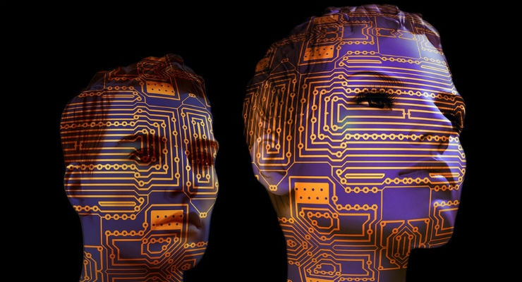


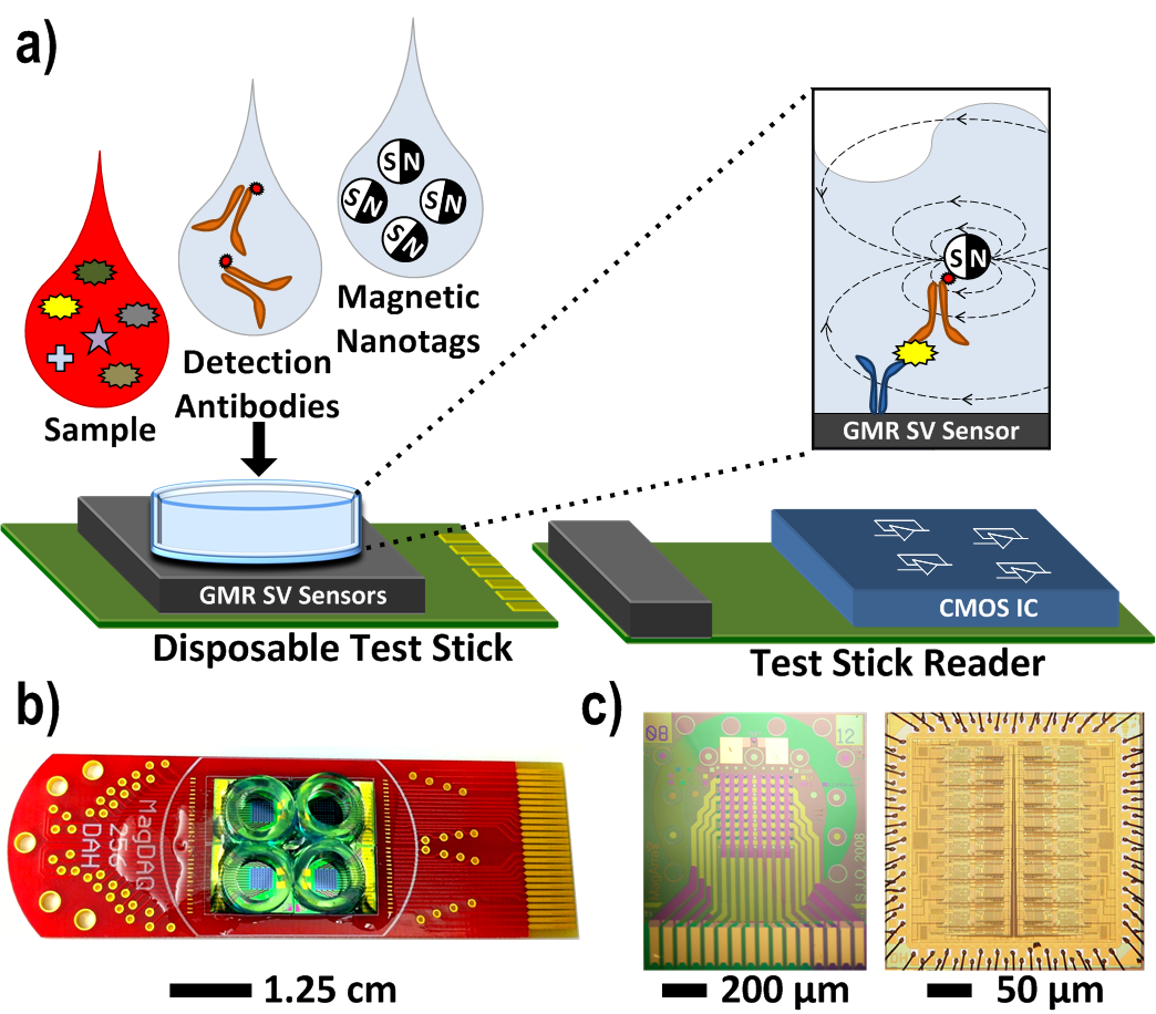
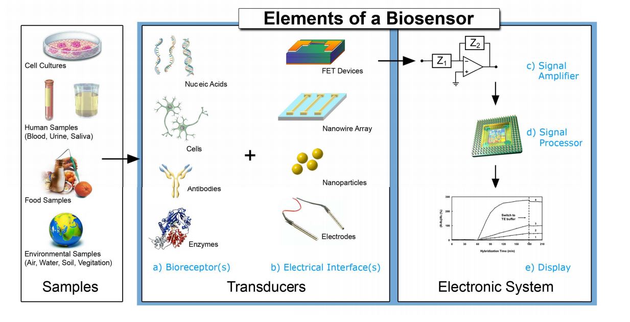


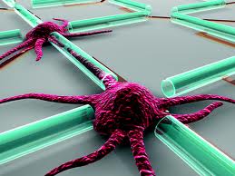


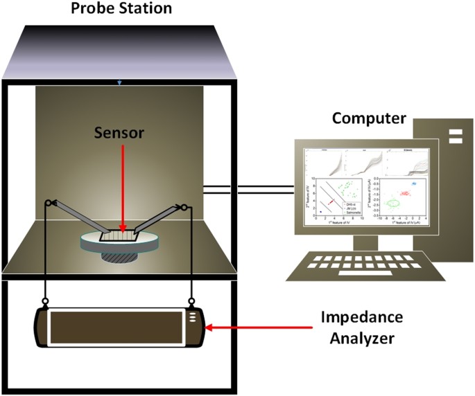


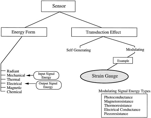
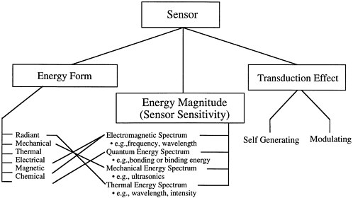
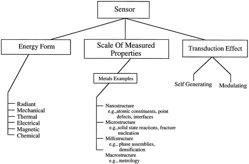
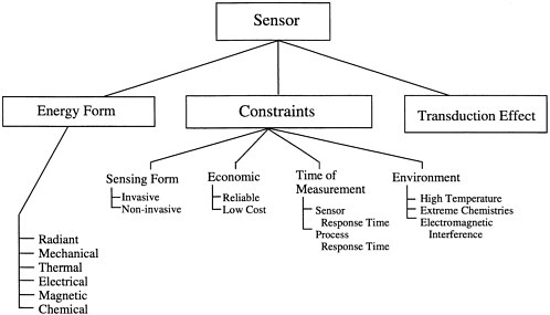


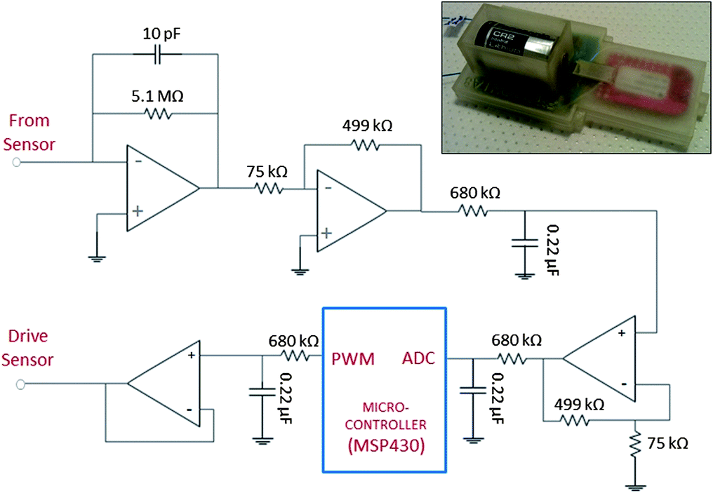
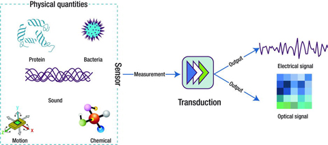
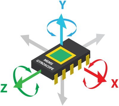
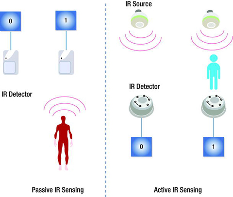
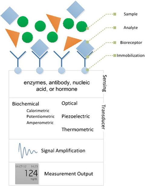

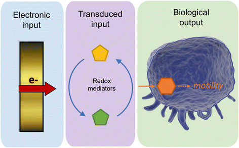

Tidak ada komentar:
Posting Komentar